Publications
Nine peer-reviewed journal articles (five as first author), a doctoral thesis, and conference contributions. Each entry opens to a plain-language summary of what the paper found. For citation counts, see Google Scholar; for identifiers, ORCID 0000-0002-9919-3812.
2026 1 article
-
 PublishedWound healingPlasma
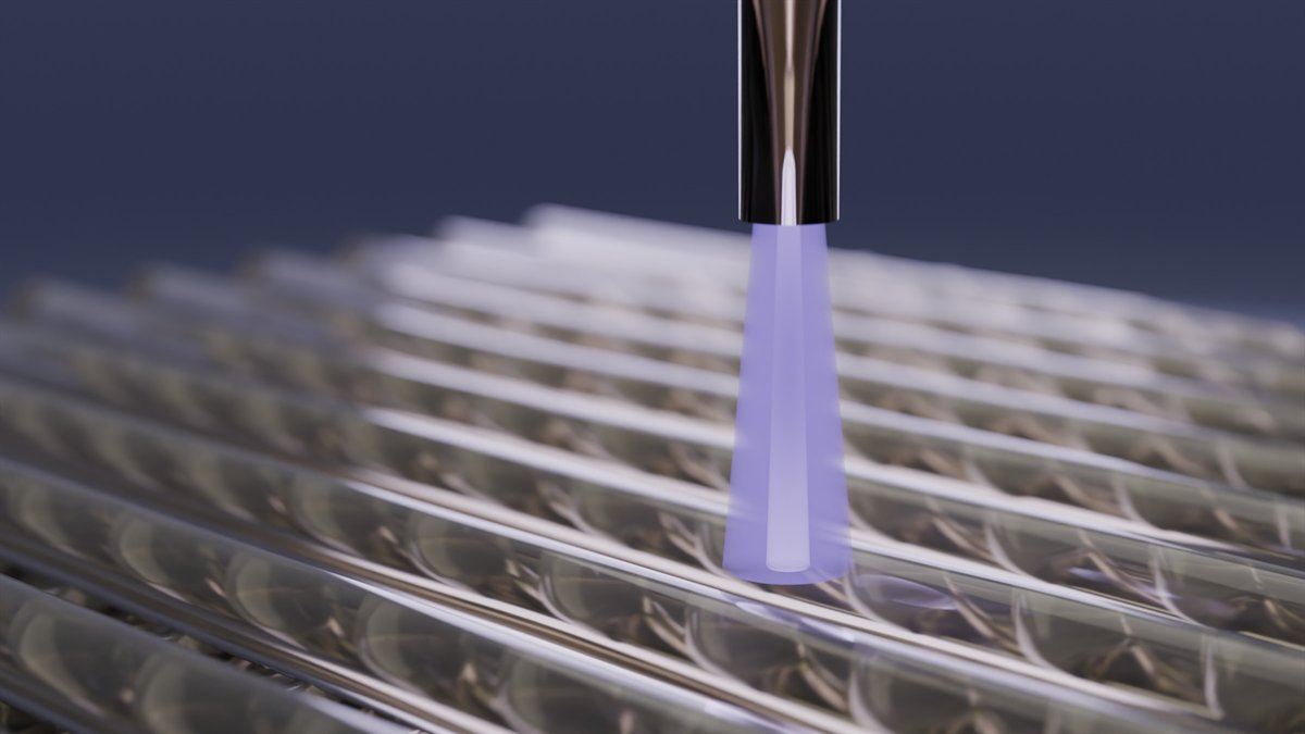
Plasma-processed bioprinted chitosan hydrogels: enhancing physicochemical and antibacterial properties for wound healing
M. Ezati, N. Mohamad, A. Hashemi, K. Polášková, D. Nečas, I. Zumberg, V. Čmiel, N. Souawda, J. Schäfer, L. Zajíčková. Plasma Processes and Polymers 23(3), e70151 (2026). doi:10.1002/ppap.70151
What did we find?
Chitosan scaffolds were bioprinted for wound care and treated with cold atmospheric-pressure plasma jets in two ways: plasma-treating the solvent before making the gel, or treating the printed scaffolds. Treating the solvent made scaffolds stronger, more heat-stable, and more antibacterial, cutting bacterial numbers at least 100-fold. Treating the finished scaffolds made their surface more bioactive, so cells attached and multiplied better.
2025 4 articles
-
 PublishedFirst authorBone
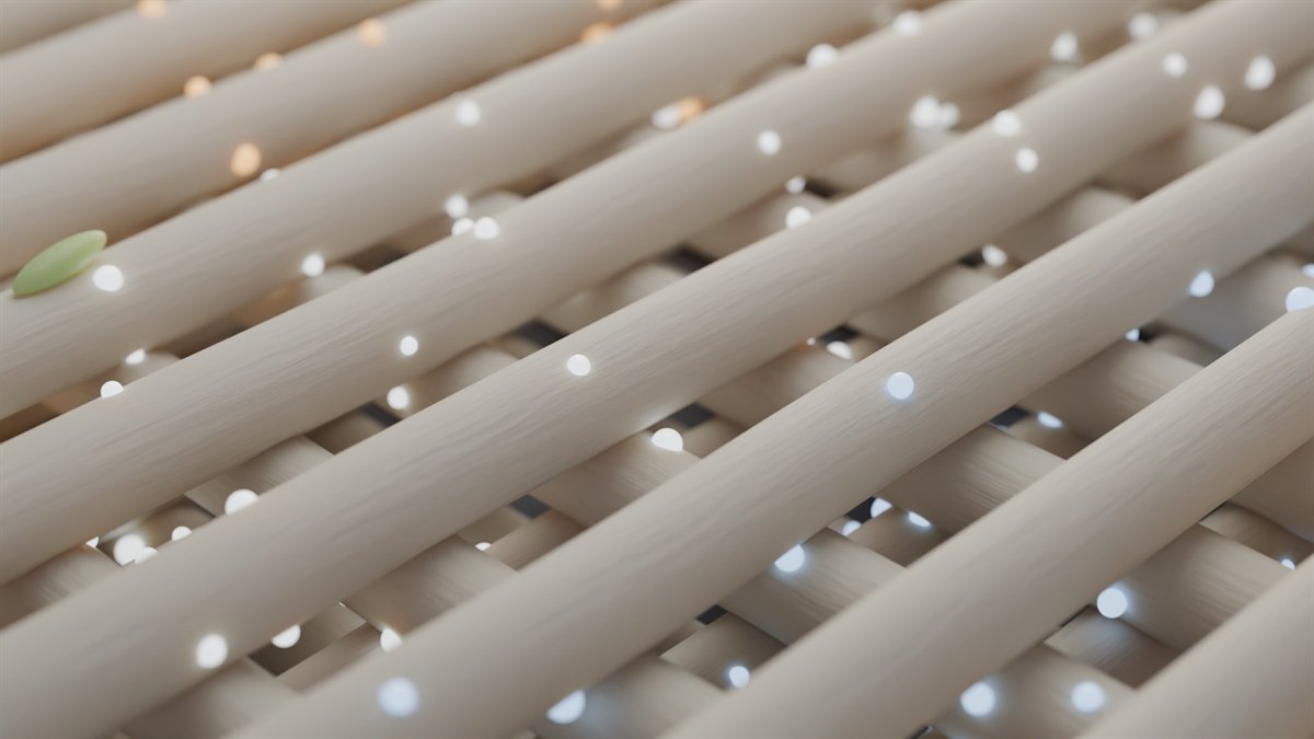
Comparative effects of ZnO, MgO, and CaO nanoparticles in 3D-printed chitosan–agarose scaffolds on antibacterial and osteogenic outcomes
A. Hashemi, M. Ezati, R. Paul, I. Zumberg, J. Bacovsky, Z. Fohlerova, V. Provaznik. Macromolecular Bioscience 25(12), e00232 (2025). doi:10.1002/mabi.202500232
What did we find?
Chitosan–agarose inks were printed into bone scaffolds loaded with zinc, magnesium, or calcium oxide nanoparticles and tested for printing, degradation, activity against E. coli and S. aureus, and effects on bone-marrow stem cells. A blend of 3.5% chitosan and 1.5% agarose printed best. Zinc oxide stood out, boosting antibacterial activity while supporting stem-cell growth and bone-type differentiation; magnesium oxide supported less cell growth, and calcium oxide was the weakest against bacteria.
-
 PublishedFirst authorHeartExosomes
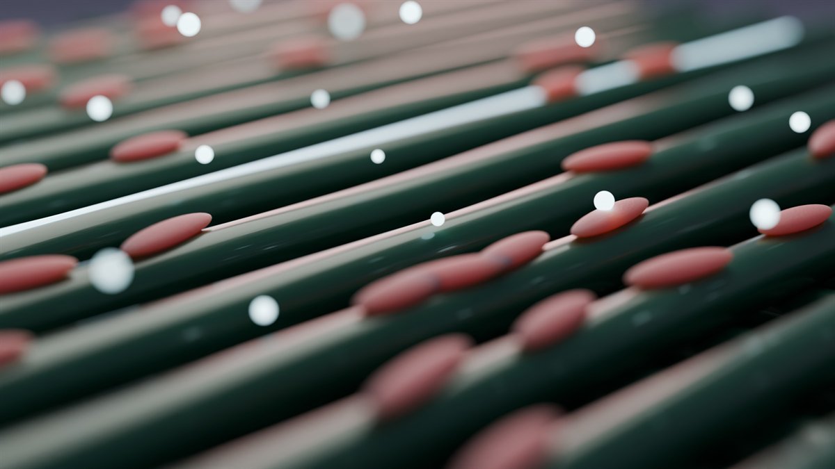
In vitro evaluation of 3D-printed conductive chitosan–polyaniline scaffolds with exosome release for enhanced angiogenesis and cardiomyocyte protection
A. Hashemi, M. Ezati, I. Zumberg, L. Chmelíková, Z. Fohlerová, V. Provazník. RSC Advances 15(21), 16826–16844 (2025). doi:10.1039/D5RA02940F
What did we find?
A printed chitosan–polyaniline scaffold carrying stem-cell exosomes was tested in a lab model of heart-attack injury, in which HL-1 heart cells were deprived of oxygen and then reoxygenated. Polyaniline raised the scaffold's conductivity, helping the heart cells survive and stay connected under low oxygen, while the exosomes boosted endothelial migration and tube formation. Together they protected heart cells and promoted new vessel growth in vitro.
-
 PublishedCancer cellsDrugs
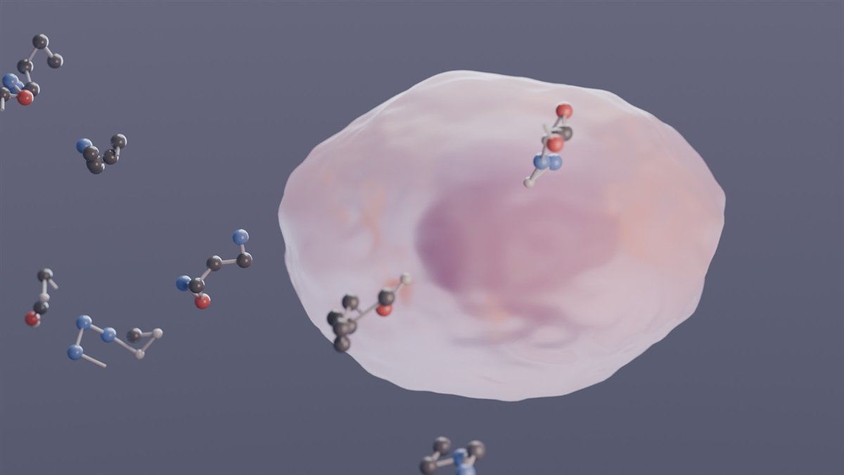
Ropivacaine and lidocaine inhibit SaOS-2 osteosarcoma cell proliferation and invasion via mitochondrial dysfunction and apoptosis
I. Zumberg, A. Hashemi, M. Ezati, L. Chmelikova, K. Ingrova, V. Cmiel. European Journal of Cell Biology 104(3), 151501 (2025). doi:10.1016/j.ejcb.2025.151501
What did we find?
Two widely used local anesthetics, ropivacaine and lidocaine, were tested on SaOS-2 human osteosarcoma cells. Both damaged the cells' mitochondria, switched on markers of programmed cell death, and reduced the cells' survival, growth, migration, and invasion, which makes them worth further study as possible add-on treatments for bone cancer.
-
 PublishedWound healingExosomes
In vitro assessment of chitosan-PEG hydrogels enriched with MSCs-exosomes for enhancing wound healing
M. Ezati, A. Hashemi, I. Zumberg, M. Partovi Nasr, Z. Fohlerova. Macromolecular Bioscience 25(5), 2400609 (2025). doi:10.1002/mabi.202400609
What did we find?
A wound dressing was made by blending chitosan and polyethylene glycol into a hydrogel carrying exosomes from fat-derived mesenchymal stem cells, releasing them steadily as the gel breaks down. In lab tests it strongly increased endothelial cell migration, lowered oxidative stress, promoted blood-vessel formation, and significantly switched on key healing-related genes.
2024 2 articles
-
 PublishedFirst authorBioinksMachine learning
Characterization and optimization of a biomaterial ink aided by machine learning-assisted parameter suggestion
A. Hashemi, M. Ezati, I. Zumberg, T. Vicar, L. Chmelikova, V. Cmiel, V. Provaznik. Materials Today Communications 40, 109777 (2024). doi:10.1016/j.mtcomm.2024.109777
What did we find?
Machine learning guided the design of a chitosan–gelatin–agarose ink for extrusion bioprinting, which was then tested for printability, flow behaviour, water affinity, degradation, and its effect on bone-marrow stem cells. The optimized ink had a viscosity suited to reliable printing, printed structures kept their shape, and the cells stayed viable with healthy morphology. The best formulation was 27% agarose, 53% chitosan, and 20% gelatin.
-
 ReviewFirst authorVesicles
Extracellular vesicles and hydrogels: an innovative approach to tissue regeneration
A. Hashemi, M. Ezati, M. Partovi Nasr, I. Zumberg, V. Provaznik. ACS Omega 9(6), 6184–6218 (2024). doi:10.1021/acsomega.3c08280
What does it argue?
Extracellular vesicles are tiny cell-released packages of proteins, lipids, and nucleic acids that can steer other cells toward repair. The review covers where they come from, how they are characterized (including exosome profiling and machine learning), which hydrogel properties make good carriers, and recent regeneration studies. Its central point: vesicles alone are cleared quickly, and hydrogels can hold them at the injury site for localized, controlled release.
2019–2020 2 articles
-
 PublishedFirst authorImplantsDrug release
Chitosan coating of TiO₂ nanotube arrays for improved metformin release and osteoblast differentiation
A. Hashemi, M. Ezati, J. Mohammadnejad, B. Houshmand, S. Faghihi. International Journal of Nanomedicine 15, 4471–4481 (2020). doi:10.2147/IJN.S248927
What did we find?
Titanium surfaces were covered with TiO₂ nanotube arrays, filled with metformin, and spin-coated with a thin chitosan layer, then tested with bone-marrow mesenchymal stem cells. Compared with uncoated nanotubes, the coated surfaces improved cell attachment, growth, and bone-differentiation markers. The coating stretched drug release from 3 days to 20 days and greatly reduced the initial burst.
-
 PublishedImplants
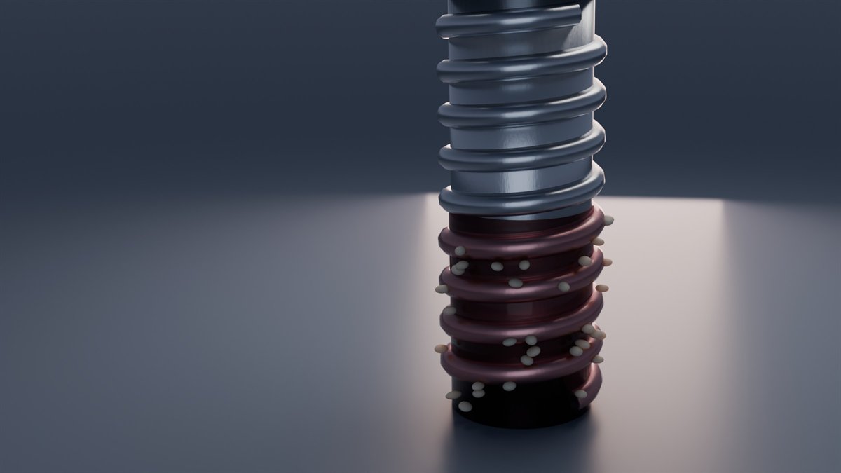
The effect of bio-conditioning of titanium implants for enhancing osteogenic activity
M. Montazeri, A. Hashemi, B. Houshmand, S. Faghihi. Journal of Oral Implantology 45(3), 187–195 (2019). doi:10.1563/aaid-joi-D-18-00020
What did we find?
Titanium discs and commercial implants were spin-coated with blood, fetal bovine serum, or saline and seeded with MG63 bone-forming cells. The coatings raised surface roughness and wettability, most of all with blood. Blood-coated titanium gave the best cell attachment and growth, closely followed by serum, and coated samples showed higher alkaline phosphatase, a marker of bone-forming activity.
Theses and conference contributions
-
 PhD thesis2025
4D bioprinting based on the maturation of engineered tissue constructs
A. Hashemi. Doctoral thesis, Brno University of Technology (defended 24 June 2025). Supervisor: Prof. Valentyna Provazník.
What is it about?
How additive manufacturing can build tissue constructs, as cell-coated scaffolds, that serve as vascular replacements or help regenerate damaged heart tissue, and how those constructs mature after printing.
-
MSc thesis2018
Deposition and functionalization of nanostructures on the surface of titanium substrates for biomedical applications
A. Hashemi. Master's thesis, University of Tehran (2018). Supervisor: Prof. Javad Mohammadnejad.
-
Poster2024
Enhancing wound healing with bioprinted chitosan hydrogels treated by cold atmospheric plasma jets
N. Mohamad, M. Ezati, A. Hashemi, K. Polášková, I. Zumberg, V. Čmiel, J. Schäfer, L. Zajíčková. ICPM10 & IWPCT9, Ljubljana, Slovenia, September 2024. Book of Abstracts.
What did we find?
Early results from the plasma project: plasma-treated printed chitosan scaffolds became more water-attracting (lower contact angle) and significantly improved fibroblast shape and growth, pointing to use as wound dressings.
-
Oral presentation2022
Machine learning–based optimization for novel bioink development
A. Hashemi (presenting author). IUPESM World Congress on Medical Physics & Biomedical Engineering (WC2022), Singapore (virtual), 12–17 June 2022.